Motor effects of clostridial neurotoxins in the central nervous system (MEFCLO)
HRZZ Installation Research Project UIP-2019-04-8277
Project leader (PI): Ivica Matak, Research Associate
Host institution: University of Zagreb School of Medicine, Zagreb, Croatia
Project duration: 60 months (01.01.2020 – 31.12.2024)
Project funding: 1.500.000,00 HRK
This project is supported by Croatian Science Foundation (HRZZ) Project call: “Installation research projects (UIP-2019-04),
PROJECT TEAM MEMBERS AND COLLABORATORS
Project team members
Ivica Matak, Research Associate, PhD (Department of Pharmacology, University of Zagreb School of Medicine, Zagreb, Croatia)
Petra Šoštarić Mužić, PhD candidate, dr. med. vet. (Department of Pharmacology, University of Zagreb School of Medicine, Zagreb, Croatia)
Višnja Drinovac Vlah, PhD, (Department of Pharmacology, University of Zagreb School of Pharmacy)
Patrik Meglić, PhD candidate, dr. med. vet. (Department of Pharmacology, University of Zagreb School of Medicine, Zagreb, Croatia)
Dalia Nemanić, MPharm, (Department of Pharmacology, University of Zagreb School of Pharmacy and Biochemistry, Zagreb, Croatia)
Collaborators and their roles:
Prof. Lidija Bach-Rojecky, PhD Department of Pharmacology, University of Zagreb School of Pharmacy and Biochemistry. Advices related to project coordination and implementation.
Prof. Zdravko Lacković, MD, PhD, the head and founder of Laboratory of Molecular Neuropharmacology (retired), University of Zagreb School of Medicine. Advices related to project coordination and implementation.
Dr. sc. Nikola Habek (MD) (Laboratory of Neurophysiology and “whole cell patch-clamp”, School of Medicine, University of Zagreb), advisor on introducing an experimental setup for electromyography, co-operation on electrophysiological experiments on cerebral cuts (not included in objectives and activities for implementation due to high risk for implementation).
Prof. dr. sc. Mario Cifrek and doc. dr.sc. Željka Lučev Vasić (Department of Electronic Systems and Information Processing, Faculty of Electrical Engineering and Computing, University of Zagreb), advices on introducing an experimental setup for electromyography, possible collaboration on joint setup construction and further collaboration.
External collaborators:
assist. prof. Ornella Rossetto, PhD; Marco Pirazzini, PhD, Research associate; Federico Fabris, M.Biotechnol / PhD candidate. Investigation of central effects of botulinum toxin serotypes other than type A botulinum toxin.
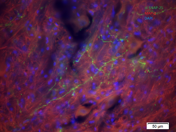
Botulinum toxin type A enzymatic activity (cleaved SNAP-25, green) in ventral horn following Its injection into the rat calf muscle.
PROJECT SUMMARY
Clostridial neurotoxins such as tetanus toxins (TeNT) and botulinum toxins serotypes A-G (BoNT/A-BoNT/G) are one of the most potent biological toxins, and causative agents of tetanus and botulism. Low therapeutic doses of BoNT/A injected into local muscles (in order of picograms to few nanograms) are used to treat hyperkinetic movement disorders and spasticity. Our preliminary studies indicate that the important part of its effect on abnormally increased muscle tone might occur in the central nervous system (CNS). Proposed project involves characterization of new preclinical models of hyperkinetic movement disorders, and characterization of the central effects of BoNT/A. The aim of present studies is to induce the imbalance of excitatory and inhibitory neurotransmission of motor regions with very low doses of TeNT and BoNT/A applied into the peripheral muscle, or into certain regions of the central nervous system. This w ill imitate hyperkinesia and hypertonia present in spasticity or dystonia. In animals w ith hyperkinesia, the effects of botulinum toxins will be characterized in extrafusal and intrafusal motor fibers, as well as central neurons of the ventral horn and brain stem. Central activity of BoNT/A will be prevented by employing intrathecally applied BoNT/A-neutralizing anti-toxin. Behavioral 2D and 3D video analysis of walking in normal and hyperkinetic animals, as well as motor tests of balance, fatigue, and the joint resistance to passive movement will be used to study the BoNT/A effects. The toxin effects will also be characterized by electromyography of spinal reflexes, and analysis of expression of neurotransmitter and proteins involved in motor neurotransmission. In the research fields of Neurology and Neuroscience, the expected scientific results are expected to advance the preclinical study of new therapeutic approaches to the treatment of movement disorders, the basic knowledge about the plasticity of the motor system, as well as the mechanisms of action of clostridial neurotoxins in the CNS.
PROJECT BACKGROUND
The project is focusing on the research of effect of clostridial neurotoxins in motor nervous system, and attempts to develop new experimental models of hyperkinetic movement disorders such as dystonia and spasticity. These disorders are characterized by prolonged or intermittent unwanted muscular contractions that cause unwanted movements and abnormal position of the body and limbs. They are common movement disorders, with great influence on the quality of life. Up to now, the most important pharmacological treatment of dystonia and one of the most important therapeutic options for spasticity are botulinum toxin type A (BoNT/A) injections into hyperactive individual muscles or muscle groups. It is considered that one of the most important therapeutic effects of botulinum toxins is the blockage of cholinergic neuromuscular transmission leading to flaccid paralysis. However, the effects of botulinum toxin can hardly be explained solely by such action. In humans it was shown that botulinum toxin can act on activity or the excitability of motoneurons that innervate distant muscle groups. The time of onset or occurrence of the peak beneficial effect in the symptoms reduction does not accompany the time course of muscle flaccid paralysis. It has been suggested that, besides the neuromuscular junctions, BoNT/A could also act on the spinal reflex system involving gamma motor neurons and Ia muscular adherents that innervate the muscular spindle, and Ib muscular afferents innervating the Golgi’s tendon organ. However, the role of action on stretch reflex elements cannot explain all the effects of botulinum toxins, especially the effects at distant muscles or muscle groups (that cannot be explained by the toxin diffusion), and the duration of useful effect which often outlasts the duration of muscle weakness caused by primary flaccid paralysis.
-In our previous studies, by examining the BoNT/A enzymatic activity in the CNS, we found that low doses of botulinum toxin, depending on axonal transport, are transported via peripheral nerves to the ventral horn of the spinal cord, specifically to cholinergic neurons surrounding the motor neurons. Our recent study has shown that the administration of BoNT/A – specific antitoxin into the cranial cerebrospinal space reduces the toxin’s enzyme activity in the facial nucleus when the toxin was applied to facial whisker pad muscles. These observations have demonstrated that the enzyme activity of BoNT/A in the motor part of the CNS should take place in the second order synapses by the trans-synaptic transport process. However, the role of botulinum toxin activity in the CNS on normal or hyperactive muscular tone, and the therapeutic role of such effect in the treatment of movement disorders is currently unknown. Our findings indicate that botulinum toxin type A centrally transported directly from the injected nerve reduces muscle hypertonia induced by intramuscular tetanus toxin administration (Matak, 2020). These findings indicate that an important part of its effect on abnormally increased muscle tone may occur in the central nervous system. This finding suggests that botulinum toxin in the CS may act on the imbalance between excitation and inhibition of transmission at different levels of the CNS. This disbalance is also the main mechanism for the development of muscular tone and hyperactivity disorders in many movement disorders such as spasticity and dystonia.
AIMS AND EXPECTED IMPACT OF THE PROJECT
– The main expected results of the project are to identify the main therapeutic sites of BoNT/A action after peripheral application in the muscle, to identify possible central actions on individual behavioral and electrophysiological parameters as well as muscle atrophy, in order to provide a more complete picture of the mode of action of BoNT/A and possible side effects after therapeutic application.
– Incorporating new animal models of movement disorders that better represent the basic etiopathogenesis of neurological disorders characterized by muscular hyperactivity. The research activities included in the project will lead to the design of new animal models of muscular hyperactivity based on the modeling of neuronal hyperactivity in the various motor regions of the CNS and their behavioral and biochemical characterization.
– Elucidating the mechanisms of existing effective medicines using new models, with the possibility of identifying new targets for drugs (receptors and synapses in the CNS that they might act upon). In animal models of muscular hyperactivity, the effect of anticholinergics (botulinum toxins, antimuscarinic drugs) will be investigated, whose mechanism of action is currently unclear.
PROJECT RESULTS
Toxins 2021 virtual meeting, held on 16th of January 2021 (Accepted abstract)
LONG-TERM EFFECTS OF BOTULINUM NEUROTOXIN TYPE A ON EXPERIMENTAL MUSCLE HYPERTONIA
Ivica Matak*, Petra Šoštarić.
Laboratory of Molecular Neuropharmacology, Department of Pharmacology, University of Zagreb School of Medicine, Zagreb, Croatia
*Corresponding author: Laboratory of Molecular Neuropharmacology, Department of Pharmacology, University of Zagreb School of Medicine, Zagreb, Salata 11, 10000 Zagreb, Croatia.
E-mail address: ivica.matak@mef.hr (I. Matak).
Introduction and Objectives: Long-term effects of botulinum neurotoxin type A (BoNT-A) are the basis of its beneficial effects on neurological disorders characterized by muscle hyperactivity, such as spasticity and dystonias. Clinical reports suggest that BoNT-A–mediated normalization of muscle hypertonia does not necessarily correlate with or outlast the duration of muscular flaccid paralysis. The present aim was to reassess these clinical observations in a preclinical model of muscle hypertonia by characterizing the duration of toxin anti-spastic activity in relation to the duration of its muscular effects.
Methods: In male Wistar rats, the muscle hypertonia was evoked by tetanus neurotoxin (TeNT) injection into the gastrocnemius (1.75-2 ng total dose). After the development of spastic hypertonia, BoNT-A was similarly injected into the ipsilateral gastrocnemius muscle 7 days post-TeNT (1, 2 and 5 U/kg//20 µL). The effects of TeNT and BoNT-A were further examined by measurements of resistance to passive ankle dorsiflexion, digit abduction score (DAS), and Basso-Beattie-Bresnahan (BBB) Locomotor Rating Scale. Muscle atrophy was quantified by measurement of the medio-lateral calf diameter. After complete recovery of BoNT-A–mediated peripheral flaccid paralysis, the animals were re-injected with TeNT (day 49 after BoNT-A injection) and assessed behaviorally for another 19 days.
Results: In the immediate period following the first TeNT injection, all BoNT-A doses employed exhibited a similar reduction of the muscle hypertonia. The TeNT-evoked muscle hypertonia ended by day 28 post-TeNT, while the remaining BoNT-A–mediated flaccid paralysis recovered in a dose-dependent manner by day 42 post–BoNT-A. After the second TeNT injection, the anti-spastic activity of BoNT-A was still present up to day 68; however, it was less prominent in rats injected with the lowest BoNT-A (1 U/kg) dose. The level of persisting muscle atrophy, which did not show signs of recovery by the end of the experiment, was similar at all doses employed.
Conclusions: In line with clinical reports, we found that the antispastic effect of BoNT-A in rats persists after the lower limb recovery from flaccid paralysis in a dose-dependent manner. Long-term actions of BoNT-A on muscle hypertonia might be mediated by a mechanism distinct from its local muscular paralytic action.
Funding: Croatian Science Foundation (project ID: UIP-2019-04-8277)
Keywords: Botulinum neurotoxin type A; Flaccid paralysis; Muscle atrophy;
Muscle hypertonia; Tetanus neurotoxin
ISCM 2022 17th International Symposium on Cholinergic Mechanisms 8 – 12 May 2022 – Dubrovnik – Croatia (Accepted abstracts)
LONG-TERM MOTOR CENTRAL EFFECTS OF BOTULINUM TOXIN TYPE A IN RATS
Petra Šoštarić1, Magdalena Matić1, Ivica Matak1
1Laboratory of Molecular Neuropharmacology, Department of Pharmacology, University of Zagreb School of Medicine, Zagreb, Croatia
E-mail: petra.sostaric@mef.hr
Botulinum toxin type A (BoNT-A) is a potent presynaptic neurotoxin and a standard therapy in hyperkinetic movement disorders, presumably due to its local muscular anticholinergic effect. However, recent experimental data point to possible central effects in the CNS. The present aim was to examine the contribution of the transcytosis-dependent central toxin action on the long term muscular function recovery in rats, as well as TeNT-evoked spastic paralysis. Rats were bilaterally injected with BoNT-A into the gastrocnemius muscle (2 U/kg) or sciatic nerve (5 U/kg). The following day, the animals were injected intrathecally (i.t.) with BoNT-A-neutralizing antitoxin. On day 62, animals were injected i.m. with tetanus neurotoxin (TeNT). In different motor tests (gait ability score, digit abduction score, rota-rod, beam walking and swimming performance), i.t. antitoxin significantly accelerated the flaccid paralysis and motor performance recovery. BoNT-A reduced the lower hind-limb diameter and muscle size without significant recovery during the entire experiment, which resulted in reduction of CMAP and H reflex. The TeNT-evoked increase in muscle tone was reduced by BoNT-A dependently on its central effect. However, the H-reflex, when corrected for reduced muscle size or reduced CMAP, was not affected by the toxin treatment, suggestive of the lack of the toxin’s direct effect on monosynaptic reflex. The enzymatic activity of the toxin, examined by cleaved synaptosomal-associated protein 25 (cSNAP-25) immunohistochemistry, was still present in neuromuscular junctions and spinal cord. The central occurrence of the cSNAP-25, present in second order spinal cord cholinergic neurons, depended on the toxin’s central transcytosis.
Long term motor effects of BoNT-A both on normal motor performance (day 1-62 ), as well as the spastic paralysis (days 62-78), are influenced by the toxin’s ongoing central action mediated by retrograde transport and transcytosis. These data suggest that clinically relevant beneficial effect of BoNT-A result from toxin’s combined peripheral and central effects.
Funding: Croatian Science Foundation (project ID: UIP-2019-04-8277)
LOW DOSE TETANUS TOXIN INJECTIONS INTO THE RAT MOTOR CORTEX AND STRIATUM IMPAIR THE NARROW BEAM WALKING PERFORMANCE
Patrik Meglić1, Petra Šoštarić1, Ivica Matak1
1Laboratory of Molecular Neuropharmacology, Department of Pharmacology, University of Zagreb School of Medicine, Zagreb, Croatia
E-mail: patrik.meglic@mef.hr
Basal ganglia and motor cortex integrate the sensory proprioceptive input arriving from periphery and the planning and execution of movement. Abnormalities in the motor cortex and basal ganglia excitability play a central role in the onset of movement disorders such as dystonias and parkinsonism. Tetanus neurotoxin (TeNT) prevents the inhibitory neurotransmission in central synapses. The aim of present study was to examine the behavioral effect of neuronal disinhibition in mentioned brain regions induced by low, non-convulsive doses of TeNT.
The rats were injected unilaterally into caudate putamen or motor cortex with 0.5 ng TeNT. After 2 weeks, the injections were repeated into the aforementioned contralateral motor regions, and the effect of TeNT was assessed for another 2 weeks. Various behavioral tests were repeatedly performed to assess the effect of TeNT-induced disinhibition on normal motor performance in rats, as well as to exclude possible epileptogenic toxin action. First unilateral toxin injection exerted a short-lasting impairment in the ability to transverse a narrow beam, and with no visible effects on rota-rod, swimming, gait analysis (catwalk) and EMG. Upon the second toxin injection, the animals developed a lasting impairment in the beam walking performance while the performance in other motor tests remained unimpaired. The plantar misplacement of the hind-limb during the narrow beam traversing were more evident on the hind-paw contralateral to the injected brain region. Open field test, pre-pulse inhibition and attempted audiogenic seizure tests did not indicate the possible epileptogenic actions of TeNT.
The motor cortex or striatal disinhibition with TeNT induces subtle motor impairment in the relatively complex motor task of crossing the elevated narrow beam, requiring correct prediction of paw placement. After unilateral toxin applications, the animals appear to be able to compensate the proprioceptive motor deficit, which is then aggravated and becomes long lasting after contralateral regional disinhibition.
Funding: Croatian Science Foundation (project ID: UIP-2019-04-8277)
10th Croatian Congress of Pharmacology with International Participation and 1st Croatian Congress of Chlinical Pharmacology with International Participation 25 – 28 2022 September – Opatija – Croatia (Accepted abstracts)
EXPERIMENTAL FOCAL MUSCLE HYPERTONIA OFFERS NOVEL INSIGHTS INTO MOTOR EFFECTS OF BOTULINUM TOXIN TYPE A
Matak I.,1 Meglić P.,1 Šoštarić P.1
1Laboratory of Molecular Neuropharmacology, Department of Pharmacology, University of Zagreb School of Medicine, Zagreb, Croatia
Introduction: Transient and reversible synaptic silencing by botulinum toxin type A (BoNT-A) is the basis of its therapeutic efficacy and safe use in movement disorders. Based on timing of BoNT-A actions and importance of appropriate muscle targeting, neuromuscular paralysis is considered its main therapeutic effect. Preclinical models have mostly focused on measurement of BoNT-A neuroparalytic effects in normal animals, in contrast to the possibly more relevant models of sustained or intermittent muscle hyperactivity. Moreover, clinical and experimental studies suggested that therapeutic effects of BoNT-A are not entirely explainable by peripheral neuromuscular site of action, suggesting the need for models of muscle hyperactivity.
Results: In a series of experiments, we employed tetanus toxin (TeNT) as an elegant way to disinhibit the motoneuronal activity and evoke focal muscle hypertonia. We found that BoNT A induces simultaneous and non-mutually exclusive antispastic activity at both peripheral muscular and central spinal level. When separated from muscular action, central trans synaptic toxin action alone was effective in reducing the experimental muscle spasm. The central antispastic effect is masked by the toxin's peripheral action in the early period post injection, and becomes more prominent as the neuromuscular effect starts to weaken (2 months post BoNT-A). The toxin's activity at peripheral and central sites, as well as the duration of its beneficial antispastic action, was found to be dose-dependent.
Conclusion: Preclinical models recapitulating some of the neurophysiological mechanisms of spastic and hyperkinetic movement disorders may be more helpful at explaining the factors and mechanisms of the BoNT-A’s long term clinical actions.
Funding: Croatian Science Foundation (project ID: UIP-2019-04-8277)
LONG TERM CENTRAL EFFECTS OF BOTULINUM TOXIN TYPE A ON MUSCULAR FUNCTION AND RECOVERY IN RAT
Šoštarić P.,1 Matić M.,1 Matak I.1
1Laboratory of Molecular Neuropharmacology, Department of Pharmacology, University of Zagreb School of Medicine, Zagreb, Croatia
Introduction: Botulinum toxin type A (BoNT-A) is one of the most potent neurotoxins. Due to persuamble action on local neuromuscular terminals its widley used as therapy in different motor disorders. However, observations in clinical use points to the possible central effects. Here, after toxin's peripheral application, we examined long termed transcytosis-dependent central effects, on normal tone muscles function and recovery, as well as tetanus neurotoxin (TeNT) evoked spasticity.
Materials and methods: BoNT-A was bilaterally injected in gastrocnemius muscle (2U/kg) or sciatic nerve (5 U/kg) of the rats. The next day intrathecall (i.t.) injections of BoNT-A-neutralizing antitoxin were administered in lumbar part of spinal cord. Following the flaccid paralysis recovery, TeNT was injected unilaterally in gastrocnemius muscle on day 62.
Results: I.t. antitoxin significantly accelerated the flaccid paralysis and motor performance recovery in different motor tests (gait ability score, digit abduction score, rota-rod, beam walking and swimming performance). Dependently on it's central effects, BoNT-A reduced increased muscle tone evoked by TeNT. However, BoNT-A didn't affect H-reflex when corrected for reduced muscle size or CMAP, persuambly due to lack of direct effect on monosynaptic reflex.
The toxin enzymatic activity examined by cleaved synaptosomal-associated protein 25 (cSNAP-25) immunohistochemistry, was still present in neuromuscular junctions and spinal cord. cSNAP-25, presence in second order spinal cord cholinergic neurons, depended on the toxin’s central transcytosis.
Conclusion: Normal motor performance (day 1-62), as well as the spastic paralysis (days 62-78), are influenced by BoNT-A long-term, ongoing central action mediated by retrograde transport and transcytosis. suggesting clinically relevant beneficial effect result from combined peripheral and central BoNT-A effects.
Funding: Croatian Science Foundation (project ID: UIP-2019-04-8277)
EFFECTS OF LOW DOSE TETANUS TOXIN INJECTIONS INTO THE RAT MOTOR CORTEX AND BASAL GANGLIA ON MOTOR PERFORMANCE
Meglić P.,1 Šoštarić P.,1 Virag D.,1 Matak I.1
1Laboratory of Molecular Neuropharmacology, Department of Pharmacology, University of Zagreb School of Medicine, Zagreb, Croatia
Introduction: Abnormalities in the motor cortex (Mc) and basal ganglia excitability play a central role in movement disorders such as dystonias and parkinsonism. Tetanus neurotoxin (TeNT) prevents the inhibitory neurotransmission in central synapses. The aim of the experiments was to examine the behavioral effect of neuronal disinhibition in mentioned brain regions induced by low, non-convulsive doses of TeNT.
Materials and methods: In the first experiment rats were unilaterally injected into the caudate putamen (CPu) or Mc with 0.5 ng TeNT. The injections were repeated into the contralateral motor regions after 2 weeks, and the effect of TeNT was assessed for another 2 weeks. In the another preliminary experiment, rats were unulateraly injected with 0.5 ng of TeNT into the globus pallidus internus (GPi) and substantia nigra (SN), or SN alone. Different behavioral tests were performed repeatedly to assess the effect of TeNT induced disinhibition on motor performance, as well as to exclude possible epileptogenic action of TeNT.
Results: The Mc or CPu disinhibition with TeNT induces subtle motor impairment during the narrow beam crossing in 50% of treated animals, without signs of epileptogenic action. Animals injected into the GPi and SN, or SN alone, showed ipsilateral circling tendency in open field (50%) and swimming tests (100%).
Conclusion: TeNT injected into defined motor regions can induce motor impairments of different modalities, depending on the site of injection. While subtle motor impairment after CPu and MC disinhibition require aggravation by contralateral regional disinhibition, unilateral GPi od SN disinhibition induced more prominent motor impairment.
Funding: Croatian Science Foundation (project ID: UIP-2019-04-8277)
BraYn 2022 5th Brainstorming Research Assembly for Young Neuroscientists 28-29-30 September 2022 – Rome – Italy (Accepted abstracts)
DARE WE LOOK BEYOND MUSCULAR EFFECTS; LASTING CENTRAL ACTION OF BOTULINUM TOXIN RISING US TO POSSIBLE CLINICAL IMPLICATIONS?
Petra Šoštarić1,
Supervisor: Dr.sc. Ivica Matak
Co-authors: Magdalena Matić1, Ivica Matak1
1Laboratory of Molecular Neuropharmacology, Department of Pharmacology, University of Zagreb School of Medicine, Zagreb, Croatia
E-mail: petra.sostaric@mef.hr
Introduction: Botulinum toxin type A (BoNT-A) is a potent neurotoxin with anticholinergic effect. It is a standard therapy in various movement disorders, presumably due to action on local neuromuscular terminals. However, clinical findings and recent experimental data, points to the possible central effects. Moreover, BoNT-A non-local actions have been reported after BoNT-A treatments as effects on distant muscles located far from treated neuromuscular junctions or muscle spindle zone. Herein, after toxin's peripheral injection, the aim was to examine the contribution of the transcytosis-dependent central toxin action, on normal tone muscle function and recovery, as well as tetanus neurotoxin (TeNT) evoked spasticity
Methods: Rats were bilaterally injected with BoNT-A into the gastrocnemius muscle (2 U/kg) or sciatic nerve (5 U/kg). To stop the toxin central transcytosis, BoNT-A-neutralizing antitoxin was intrathecally (i.t.) administered after 24 hours. After recovery from flaccid paralysis, TeNT was intramuscularly (i.m.) injected to animals on day 62, and animals were followed for next 14 days, until they recovered from evoked spastic paralysis.
Results: In different motor tests (gait ability score, digit abduction score, rota-rod, beam walking, catwalk), i.t. antitoxin significantly accelerated the flaccid paralysis and motor performance recovery. TeNT-evoked increase in muscle tone was reduced by BoNT-A dependently on its central effect. However, the H-reflex, when corrected for reduced muscle size or reduced compound muscle action potential (CMAP), was not affected by the toxin treatment, suggestive of the lack of the toxin’s direct effect on monosynaptic reflex. The toxin enzymatic activity examined by cleaved synaptosomal-associated protein 25 (cSNAP-25) immunohistochemistry, was still present in neuromuscular junctions and spinal cord. cSNAP-25, presence in second order spinal cord cholinergic neurons, depended on the toxin’s central transcytosis.
Conclusion: Long term motor effects of BoNT-A both on normal motor performance (day 1-62), as well as the spastic paralysis (days 62-78), are influenced by the toxin’s ongoing central action mediated by retrograde transport and transcytosis. These data suggest that clinically relevant beneficial effect of BoNT-A result from toxin’s combined peripheral and central effects.
Funding: Croatian Science Foundation (project ID: UIP-2019-04-8277)
19TH WORLD CONGRESS OF BASIC & CLINICAL PHARMACOLOGY 2023, 2 – 7 June, 2023, Glasgow, Scotland, United Kingdom
THE TOXIN TRIO AND ONE ANTITOXIN COMBINED TOGETHER TO UNDERSTAND THE MECHANISMS OF ACTION OF BOTULINUM TOXIN TYPE A IN THE MOTOR SYSTEM
Petra Šoštarić1, Patrik Meglić1, Ivica Matak1
1Laboratory of Molecular Neuropharmacology, Department of Pharmacology, University of Zagreb School of Medicine, Zagreb, Croatia
E-mail: petra.sostaric@mef.hr
Introduction: Botulinum toxin A (BoNT-A) is nature's most potent presynaptic neurotoxin, which is used as a standard therapy in different hyperkinetic movement disorders. The therapeutic effect of BoNT-A in hyperactive muscle is classically attributed to neuroparalysis of muscular motor terminals, however, recent research revealed the toxin's enzymatic activity in the central nervous system and lasting effects on focal muscle hypertonia, at least in part due to the toxin's central transcytosis. The aim of the study was to separate the relative contribution of BoNT-A peripheral and central effects on normal and spastic muscle by employing subsequent latrotoxin (LTX) and neutralizing antitoxin to circumvent its muscular or spinal cord enzymatic action and to challenge its efficacy in muscle hypertonia by tetanus neurotoxin (TeNT). Methods: Male Wistar rats (N=6-8/group) were unilaterally injected into gastrocnemius muscle with BoNT-A (Clostridium botulinum type A neurotoxin complex, 5 U/kg, each unit corresponding to 48 pg toxin complex). After 24 hours, BoNT-A-neutralizing antitoxin (5 I.U.) was administered intrathecally to prevent the toxin's central transcytosis. LTX (4 μg) was administered in gastrocnemius on day 3 after BoNT-A to abolish the toxin's peripheral effects. Motor tests such as gait ability test, digit abduction score (DAS) and compound muscle action potential (CMAP) measurements were repeatedly performed to assess motor performance recovery. After the animals recovered from flaccid paralysis, unilateral injection of TeNT (1,5 ng) was administered to gastrocnemius to induce spastic paralysis. Recovery of the spastic paralysis was assessed by ankle dorsiflexion resistance test and monosynaptic H-reflex measurements. Results: The BoNT-A induced lasting and reversible impairment of examined motor functions, which mostly recovered by day 70 post i.m toxin injection. Destruction and fast regeneration of peripheral motor terminals by LTX accelerated the recovery of motor function and counteracted the lasting effects of BoNT-A on CMAP reduction and muscle atrophy. The TeNT increased monosynaptic H-reflex was not decreased by LTX or BoNT-A treatment. Conclusion: BoNT-A effects on atrophy, gait ability and DAS most likely depend on the peripheral action of the toxin, due to the accelerated recovery enhanced by LTX-mediated elimination of BoNT-A poisoned neuromuscular junctions and their fast subsequent regeneration. TeNT-evoked spastic paralysis was affected by BoNT-A central and peripheral effects. This data suggest that combined peripheral and central effects of the peripherally administered BoNT-A are responsible for its lasting action in hyperactive muscle, while the peripheral toxin effect is more important in motor performance impairment and lasting muscle atrophy.
EFFECTS OF LOW DOSE TETANUS TOXIN INJECTIONS INTO THE RAT BASAL GANGLIA ON MOTOR PERFORMANCE
Patrik Meglić1, Petra Šoštarić1, Ivica Matak1
1Laboratory of Molecular Neuropharmacology, Department of Pharmacology, University of Zagreb School of Medicine, Zagreb, Croatia
E-mail: patrik.meglic@mef.hr
Abstract
Introduction: Abnormalities in the basal ganglia activity play a central role in the onset of movement disorders such as dystonia and parkinsonism. The basal ganglia and related nuclei are engaged primarily in motor control, together with a wider variety of roles such as motor learning, executive functions, behavior and emotions [1.]. Tetanus toxin (TeNT) causes neuronal disinhibition by selectively blocking the inhibitory GABA- or glycinergic synapses [2.]. The aim of the present study was to examine the behavioral effect of neuronal disinhibition in the basal ganglia induced by low, non-convulsive doses of TeNT. Methods: Male Wistar rats (N=9/group) were unilaterally stereotaxicaly injected into the caudate putamen (CPu) with 0.8 ng of TeNT while smaller globus pallidus internus (GPi) and substantia nigra (SN) regions were injected with 0.4 ng of TeNT. The effects of TeNT were assessed on the 7th, 10th and 14th day after the injection. Motor and behavioral tests such as beam-walking, ladder-walking, gait analysis, open field and swimming test were repeatedly performed to assess the effect of TeNT-induced disinhibition on normal motor performance. On the 10th day after the injection, animals underwent an amphetamine-induced rotation test (D-amphetamine 1 mg/kg i.p.) to intensify ipsilateral circling visible in the open field and swimming tests. An audiogenic seizure test was performed on the last day of experiment to exclude possible epileptogenic action of TeNT. Results: After unilateral TeNT injection into the CPu, plantar misplacement of the hind-limb during the beam-walk test was evident on the hind-paw contralateral to the injection site. Animals injected into the CPu, GPi and SN showed a tendency to ipsilateral circling behaviour in the open field and swimming tests, which was even more emphasized during the amphetamine- induced rotation test. Conclusion: Disinhibition of CPu with TeNT induces visible contralateral motor impairment in beam-walk test, a relatively complex motor task requiring correct prediction of hind-paw placement. On the other hand, disinhibition of all three injected brain regions induced an ipsilateral circling tendency. Later result, reminiscent of the effects of experimental parkinsonism-inducing dopaminergic nerotoxins in combination with D-amphetamine, suggests important interaction of inhibitory and dopaminergic transmission in basal ganglia for extrapyramidal motor control and impairment.
BEYOND TRADITIONAL MUSCULAR APPLICATION: TARGETING MULTIPLE MOTOR UNITS AND LOWER LEG MOTOR FUNCTIONS BY INTRA-SCIATIC BOTULINUM TOXIN INJECTIONS IN RATS
Ivica Matak1, Petra Šoštarić1, Magdalena Matić1
1Laboratory of Molecular Neuropharmacology, Department of Pharmacology, University of Zagreb School of Medicine, Zagreb, Croatia
E-mail: ivica.matak@mef.hr
Classically, targeting different muscles by multiple injection sites is assumed to be the only feasible mode of botulinum toxin type A (BoNT-A) injection in affected spastic or dystonic limb/region. However, recent preclinical studies demonstrated the role of BoNT-A axonal transport and central transcytosis in muscle spasm relief. Thus, we examined if targeting the common peripheral nerve more proximally to the CNS might, due to axonal traffic of the toxin, enable simultaneous targeting of different muscles at peripheral and spinal synaptic sites relevant for their neuromotor control. Method: In adult Wistar male rats (N=8/group), we investigated the effects of low dose BoNT-A bilaterally injected into the sciatic nerve trunk at mid-thigh level (INN: Clostridium botulinum type A neurotoxin complex, 2 international units or 96 pg per nerve) on lower leg motor functions (digit abduction, gait, balance, fatigue, swimming) for up to day 56 post BoNT-A. Later we examined its effect on focal muscle hyperactivity & increased monosynaptic reflex evoked by subsequent tetanus toxin injection into the gastrocnemius (TeNT, 1.5 ng, days 62-78 post BoNT-A). To assess the contribution of central sites of action, we assessed the toxin effect in combination with intrathecally delivered BoNT-A neutralizing antitoxin (5 i.u.). By employing immunodetection of cleaved synaptic target of BoNT-A, we examined the toxin's enzymatic action in tibialis anterior, gastrocnemius, and lumbar spinal cord. Results: From the nerve, BoNT-A was anterogradely transported into the corresponding lower leg muscle motor terminals, and retrogradely into the lumbar ventral horn. Combined peripheral and transcytosis-dependent central toxin actions induced longer lasting and more intensive reversible motor impairments versus its isolated peripheral effect. Similarly, the late antispastic effects of BoNT-A on TeNT-evoked focal muscle hypertonia were dependent on the toxin activity in propriospinal second-order synapses, but not the synaptic terminals mediating the monosynaptic H-reflex. Independently from its central action, peripheral toxin effect resulted in non-recovering muscle atrophy. The toxin entered the nerve even if applied briefly on the surface of isolated undamaged nerve trunk, suggesting a high affinity mode of entry through axons. Conclusions: Newly discovered BoNT-A anterograde and retrograde trafficking in primary motor neurons propose the need for investigation of non-canonical modes and sites of toxin action on neuromotor control for a therapeutically desirable effect in movement disorders. When injection into multiple muscular sites is not feasible, toxin application by existing regional nerve block techniques might provide an alternative way to target regional muscle groups.
COMPLEX MECHANISMS OF BOTULINUM TOXIN TYPE A ACTION ON PAIN AT THE LEVEL OF THE FIRST SYNAPSE
Dalia Vađunec1, Lidija Bach-Rojecky1, Ivica Matak2
1Department of Pharmacology, University of Zagreb, Faculty of Pharmacy and Biochemistry, Domagojeva 2, Zagreb, Croatia,
2Department of Pharmacology, University of Zagreb, School of Medicine, Šalata 11, Zagreb, Croatia
Email:daliavad@gmail.com
Introduction: Botulinum neurotoxin type A (BoNT-A) cleaves SNAP-25, a part of cellular machinery that mediates neurotransmitters exocytosis [1]. Its antinociceptive effect is mediated by complex mechanisms in the central nervous system, possibly at the level of the first synapse [2]. This study aimed to investigate antinociceptive effect of BoNT-A within the trigeminal nucleus caudalis (TNC) after its peripheral application. Methods:Wistar rats were assigned into four experimental groups (n=32, 8 rats per group). Control groups were injected with: saline unilaterally in the facial vibrissae (day 0); horse serum into cisterna magna (5 U/10µL, day 1), and saline or formalin 2.5% bilaterally in the vibrissae pad, (day 7). Third and fourth group (experimental groups 1 and 2) were treated as follows: BoNT-A (7 IU/kg) was injected in the vibrissae pad unilaterally (day 0), horse serum or antitoxin for BoNT-A (2 U/10µL) were injected into the cisterna magna (day 1), and formalin 2.5% was injected bilaterally into the vibrissae pad (day 7). The time rats spent rubbing the formalin-injected sites was measured for 45 minutes. Immunohistochemical analyses of c-Fos (a marker of neural activation) and cleaved SNAP-25 (a marker of BoNT-A activity) were performed in the sections of the TNC. Three sections per animal were analyzed. Statistical analysis was performed with two-way ANOVA and post-hoc Bonferroni test with p < 0.05 considered as significant. Experimental procedures were conducted in accordance with the European Union Directive 2010/63/EU and approved by the institutional Ethics Committee. Results: Peripheral BoNT-A reduced the time that rats spent rubbing the formalin-injected area in the inflammatory phase of the test (time point 2), while antitoxin prevented this effect. Immunohistochemical data showed that BoNT-A reduced bilateral expression of c-Fos positive neurons, while the antitoxin reduced this effect ispilaterally. Antitoxin significantly reduced the BoNT-A associated expression of cleaved SNAP-25 at the ipsilateral side of the TNC (Table 1). Ipsilateral represents the BoNT-A application side, while contralateral indicates BoNT-A un-injected side.
IBRO World Congress of Neuroscience 2023, 09.-13.09.2023, Granada, Spain
LOW DOSE TETANUS TOXIN INJECTIONS INTO THE RAT BASAL GANGLIA IMPAIR MOTOR PERFORMANCE
October 2023, IBRO Neuroscience Reports, 15:S697-S6
DOI:10.1016/j.ibneur.2023.08.1409
Patrik Meglić1, Petra Šoštarić1, Ivica Matak1
1Laboratory of Molecular Neuropharmacology, Department of Pharmacology, University of Zagreb School of Medicine, Zagreb, Croatia
E-mail: patrik.meglic@mef.hr
Abstract:
Abnormalities in the basal ganglia activity play a central role in the onset of movement disorders such as dystonia and parkinsonism. The basal ganglia and related nuclei are engaged primarily in motor control. Tetanus toxin (TeNT) causes neuronal disinhibition by selectively blocking inhibitory GABA or glycinergic synapses. The aim of the present study was to examine the behavioral effects of neuronal disinhibition in the basal ganglia induced by low, non-convulsive doses of TeNT. Male Wistar rats were unilaterally stereotaxicaly injected with tetanus toxin into the caudate putamen (CPu), globus pallidus internus (GPi) and substantia nigra (SN). The effects of TeNT were assessed on the 7th, 10th and 14th day after the injection. Different motor and behavioral tests were repeatedly performed to assess the effect of TeNT-induced disinhibition on normal motor performance. On the 10th day after the injection, animals underwent an amphetamine-induced rotation test. After unilateral TeNT injection into the CPu, plantar misplacement of the hind limb during the beam-walk test was evident on the hind-paw contralateral to the injection site. Animals injected into the CPu and GPi showed a tendency to ipsilateral circling behavior in the open field and swimming tests, which was even more emphasized during the amphetamine-induced rotation test. CPu and GPi injected animals displayed higher stride frequency and shorter swing time on the CatWalk analysis. GPi and CPu injected animals exhibit similar ipsi-rotational behavior and similar gait related impairments while only CPu injected animals show a proprioceptive deficit. Some results are reminiscent of the effects of experimental parkinsonism-inducing dopaminergic nerotoxins in combination with D-amphetamine. This may suggest an important interaction of inhibitory and dopaminergic transmission in the basal ganglia for extrapyramidal motor control and impairment. It also opens up a question: does TeNT suppress dopaminergic transmission in the basal ganglia?
INVESTIGATING THE LONG-LASTING EFFECTS OF BOTULINUM TOXIN TYPE A ON MOTOR PERFORMANCE AND ATROPHY
October 2023 IBRO Neuroscience Reports 15:S717-S718
DOI: 10.1016/j.ibneur.2023.08.1458
Petra Šoštarić1, Patrik Meglić1, Ivica Matak1
1Laboratory of Molecular Neuropharmacology, Department of Pharmacology, University of Zagreb School of Medicine, Zagreb, Croatia
E-mail: petra.sostaric@mef.hr
Abstract:
Botulinum toxin A (BoNT-A) is nature’s most potent presynaptic neurotoxin, which is used as standard therapy in different hyperkinetic movement disorders. Therapeutic effect of BoNT-A in hyperactive muscle is classically attributed to peripheral neuroparalysis of neuromuscular terminals, however, recent research revealed the toxin's enzymatic activity in the central nervous system and lasting effects on focal muscle hypertonia, at least in part due to the toxin's central transcytosis. The aim of the study was to separate the relative contribution of BoNT-A peripheral and central effects on normal and spastic muscle by employing subsequent latrotoxin (LTX) and neutralizing antitoxin to circumvent its muscular or spinal cord enzymatic action. Male Wistar rats (N=6-8/group) were unilaterally injected into gastrocnemius muscle with BoNT-A (5U/kg). BoNT-A-neutralizing antitoxin (5 I.U.) was administered intrathecally to prevent the toxin's central transcytosis 24 post BoNT-A injections. Latrotoxin (4 μg) was administered in gastrocnemius on day 3 after BoNT-A to abolish the toxin's peripheral effects. Motor performance recovery was repeatedly assessed with different motor tests, such as, gait ability test, catwalk analysis, digit abduction score (DAS) and compound muscle action potential (CMAP) measurements until the point where animals recovered. The BoNT-A induced lasting and reversible impairment of examined motor functions, which mostly recovered by day 70 post intramuscular toxin injection. Destruction and fast regeneration of peripheral motor terminals by latrotoxin accelerated the recovery of motor function and counteracted the lasting effects of BoNT-A on CMAP reduction and muscle atrophy. BoNT-A effects on atrophy, gait ability and DAS most likely depend on the peripheral action of the toxin, due to the accelerated recovery enhanced by latrotoxin-medited elimination of BoNT-A poisoned neuromuscular junctions and their fast subsequent regeneration. This data suggest that peripheral toxin effect are more important in motor performance impairment and lasting muscle atrophy.
Toxins 2024 7th International Conference
17-20 January 2024 • Berlin, Germany
FABS FROM PURIFIED HUMABS OPEN TO THE INTRATHECAL TREATMENT OF TETANUS
https://doi.org/10.1016/j.toxicon.2024.107397
January 2024 Toxicon 237:107398
Federico Fabris a, Petra Šoštarić b, Patrik Meglić b, Davide Corti c, Antonio Lanzavecchia c, Ornella Rossetto a d e, Cesare Montecucco a e, Marco Pirazzini a d
a Department of Biomedical Sciences, University of Padova, Padova, Italy
b Department of Pharmacology, School of Medicine, University of Zagreb, Zagreb, Croatia
c Humabs BioMed SA, Bellinzona, Switzerland
d Myology Research Center, University of Padova, Padova, Italy
e CNR-Institute of Neuroscience, Padova, Italy
Tetanus neurotoxin (TeNT) is the causative agent of tetanus, a neuroparalytic disease characterized by muscle spasticity following TeNT activity in inhibitory interneurons within the spinal cord. A worldwide clinical practice is the administration of anti-TeNT immunoglobulins (TIG) used both as prophylaxis to prevent tetanus development or to treat patients already displaying tetanus symptoms. TIG neutralizes TeNT in peripheral body fluids before it enters peripheral nerves and is retro-transported to the spinal cord. More effective would be the intrathecal administration of TIG to block TeNT in its very site of action but this is limited by the amount of protein that can be safely injected into the cerebrospinal fluid. This drawback can be overcome by highly purified human monoclonal antibodies (humAbs), which are emerging as superior therapeutics against several diseases. By screening immortalized memory B cells from immunized human donors, we isolated TT104 and TT110, two humAbs that display an unprecedented neutralization ability blocking TeNT binding at the neuromuscular junction or the translocation of the light chain into the neuronal cytosol. These antibodies were tested via the intramuscular and intrathecal administration routes in experimental mouse and rat models of tetanus. The combination of TT104 and TT110 humAbs displayed prophylactic activity when injected long before TeNT, and in post-exposure prevention of tetanus when administered within 6 hours after TeNT. None of these treatments blocked TeNT when administered beyond the 12 hours after TeNT inoculation. Conversely, the TT104-Fab (fragment antigen-binding) and TT110-Fab combination was effective in both generalized and local tetanus when administered via the intrathecal route. Therefore, TT104 and TT110 humAbs and their Fab derivatives meet all requirements to improve the current prophylaxis and therapy of human tetanus.
Funding
This research was funded by the University of Padova with the “Progetto DOR 025271” (M.P.) and “Progetto DOR 205071” (O.R.), and by the Croatian Science Foundation project: HRZZ-UIP-2019-04Fs-8277.
BOTULINUM NEUROTOXIN TYPE A REGULATES SPINAL PREMOTOR INPUTS PARTICIPATING IN NORMAL LOCOMOTION AND EXPERIMENTAL SPASTICITY IN RATS
https://doi.org/10.1016/j.toxicon.2024.107453
January 2024Toxicon 237(48):107453
DOI: 10.1016/j.toxicon.2024.107453
Ivica Matak1, Petra Šoštarić1,
1Laboratory of Molecular Neuropharmacology, Department of Pharmacology, University of Zagreb School of Medicine, Zagreb, Croatia
E-mail: ivica.matak@mef.hr
Introduction Botulinum neurotoxin type A (BoNT-A) normalizes the sustained and intermittent focal muscular hyperactivity in spasticity and dystonia. A key feature of low-dose BoNT-A therapy is the long-term reversible neuromuscular blockade of injected hyperactive muscles. However, clinical observations of modified or reduced excitability of distant motoneuronal pools and changes in recurrent and reciprocal inhibition in untreated muscles indicate a more complex BoNT-A motor effect beyond locally targeted extrafusal and intrafusal terminals. Building on previous research that demonstrated the BoNT-A axonal transport and transcytosis in motor nuclei, our aim was to characterize the motor effects of BoNT-A at spinal cord level.
Methods We examined the long-term effect of low-dose intramuscular (i. m.) BoNT-A (INN: Clostridium botulinum neurotoxin type A complex; 1–5 U/kg) on experimental rat calf muscle spasm evoked by i.m. tetanus toxin (TeNT; 1.5 ng). In rats bilaterally injected with BoNT-A into sciatic nerves (2 U per nerve) we examined the toxin actions on coordinated locomotor tasks (beamwalk, rotarod), high-intensity swimming task that requires intense, sustained motoneuronal firing, and the lower leg muscle weakness (hindlimb toe abduction and weight support during stance). The contribution of the BoNT-A central action at spinal premotor inputs was assessed by spinal intrathecal BoNT-A–specific antitoxin (5–10 i.u.) aimed at neutralizing the free toxin during its central trans-synaptic transport.
Results By examining both the different motor behavioral assessments in concert with the toxin’s enzymatic activity at the peripheral neuromuscular junction and spinal cord, we found that intramuscular BoNT-A exerted a lasting, dose-dependent action in a model of experimental local spasm. The role of the toxin’s central antispastic action was found to be more prominent at a later stage (2–2.5 months post intramuscular BoNT-A) when its primary neuromuscular effect substantially recovers. In rats bilaterally injected with BoNT-A into sciatic nerves, combined peripheral and transcytosis-dependent central toxin actions reduce the normal locomotor performance in different balance and fatigue tests, swimming speed, as well as localized muscle weakness. The toxin’s central action was not associated with modulation of the monosynaptic reflex synaptic strength (assessed by H reflex/M-wave ratio), in line with lack of enzymatic SNAP-25 (synaptosome-associated protein of 25 kDa) cleavage at ventral horn type I vesicular glutamate transporter (VGlut-1)–expressing primary afferents. Comparison of ventral horn cleaved SNAP-25 colocalization with ChAT (choline acetyltransferase) versus a general synaptic marker synaptophysin and the high-affinity BoNT-A acceptor SV2C (synaptic vesicle glycoprotein 2C), indicated the toxin action in cholinergic and a broader population of noncholinergic central excitatory synapses. This is in line with the role of BoNT-A central action in a large number of motor tasks as opposed to swimming performance only (expected if cholinergic C-boutons were the primary BoNT-A target) and counteraction of TeNT-evoked local spasm (resulting from disruption of different spinal inhibitory pathways).
Conclusions Present findings demonstrate that BoNT-A affects central premotor inputs belonging to spinal locomotor circuits that regulate skilled movement. These long-term central toxin actions also participate in the lasting antispastic activity of BoNT-A. Given that purposeful movement is commonly impaired in focal spasticity and dystonias, BoNT-A–mediated control of muscle hyperactivity may involve the spinal premotor inputs participating in the normal locomotor performance.
Funding and Acknowledgement This research was funded by the Croatian Research Foundation (project no. HRZZ UIP-2019–04-8277). Lyophilized polyclonal equine IgG-based BoNT-A antitoxin (NIBSC code 14/174) and non-affinity purified rabbit polyclonal antibody to BoNT-A-cleaved SNAP-25 recognizing the SNAP-25 1-197 fragment were a kind gift from Dr. Thea Sesardic (National Institute for Biological Standards and Control, Potters Bar, United Kingdom).
TETANUS TOXIN – EVOKED DISINHIBITION OF THE RAT BASAL GANGLIA INDUCES MOTOR IMPAIRMENTS CONSISTENT WITH PARKINSONISM
January 2024Toxicon 237(12):107457
DOI: 10.1016/j.toxicon.2024.107457
Patrik Meglić1, Petra Šoštarić1, Ivica Matak1
1Laboratory of Molecular Neuropharmacology, Department of Pharmacology, University of Zagreb School of Medicine, Zagreb, Croatia
Introduction Nuclei of the basal ganglia exert a primary role in extrapyramidal motor control, but also have broader roles encompassing motor learning, executive functions, behavior, and emotions.1 Abnormalities within neuronal circuits of the basal ganglia system are key to the development of movement disorders such as dystonia and parkinsonism. In the central nervous system, tetanus toxin (TeNT) induces neuronal disinhibition by selectively blocking inhibitory synapses in neuronal projection-connected brain regions, undergoing axonal and trans-synaptic transport.2 This study aimed to investigate the behavioral impact of neuronal disinhibition in the basal ganglia and their interconnected regions, induced by low, non-convulsive doses of TeNT.
Methods Male Wistar rats (N = 9/group) were unilaterally injected in the caudate putamen (CPu) with 2 × 0.4 ng of TeNT (distributed to 2 injection sites located rostro-caudally), while smaller regions like the globus pallidus internus (GPi) and substantia nigra (SN) received single injections of 0.4 ng of TeNT. Various motor behavioral tests including beam-walking, ladder-walking, gait analysis, open field exploration, and swimming tests, were conducted repeatedly (Days 0, 7, 10, 14) to assess the effect of TeNT-induced disinhibition on normal motor performance. On the tenth day after receiving TeNT, rats were subjected to a D-amphetamine-induced rotation test (1 mg/kg i.p.), which typically induced ipsilateral circling in hemi-parkinsonism models. Finally, audiogenic seizure tests were conducted on the last day of the experiment to rule out any potential epileptogenic effects of TeNT. Brain tissue was collected for immunohistochemical staining and western blot analysis.
Results Disinhibition of CPu with TeNT induces visible hind limb contralateral motor impairment during the beam-walk test, a relatively complex motor task requiring precise hind-paw placement prediction. Animals who received injection to the CPu or GPi exhibited ipsiversive circling behavior in the open field and swimming tests. Circling behavior in the open field test was exaggerated by D-amphetamine. Animals that received injections in the CPu and GPi showed an increase in stride frequency and a decreased swing time in catwalk gait analysis. Evident recovery of motor symptoms towards the end of study suggested reversibility of TeNT-evoked impairments. No signs of epileptogenic behavior were observed in the audiogenic seizure test, while tyrosine hydroxylase regional content suggests lack of TeNT-evoked excitotoxic degeneration of dopaminergic projection within the basal ganglia. Animals injected in GPi and CPu exhibited hemiparkinsonism/parkinsonism-like motor impairments, showcasing comparable rotational behavior, gait-related issues, and a proprioceptive deficit after CPu injections. These findings underline the complex interplay between inhibitory and dopaminergic transmission in the basal ganglia, a critical factor for motor control and function.
Conclusion TeNT, primarily entering GABA-ergic neurons in supraspinal brain regions, may be used as a tool to study the intricate system of GABA-ergic circuits and projections, as well as the significant interaction of inhibitory and dopaminergic transmission within basal ganglia-shaping extrapyramidal motor regulation and function. Further investigations are needed to pinpoint the exact affected neurotransmission pathways, which could potentially shed light on the general mechanism behind parkinson-ism-like motor impairments.
Funding Croatian Science Foundation (project ID: UIP-2019-04-8277)
BOTULINUM NEUROTOXIN TYPE A ANTINOCICEPTIVE ACTIVITY IN SPINAL CORD AND TRIGEMINAL SENSORY NOCICEPTIVE NUCLEI INVOLVES CENTRAL TRANS-SYNAPTIC TRANSPORT
January 2024 Toxicon 237(12):107463
https://doi.org/10.1016/j.toxicon.2024.107463
DOI: 10.1016/j.toxicon.2024.107463
Introduction Botulinum neurotoxin type A (BoNT-A) interferes with neurotransmitter exocytosis by endoproteolysis of synaptosomal-associated protein of 25 kDa (SNAP-25).1 While its effects in peripheral cholinergic autonomic and muscular synapses are well-known,2 BoNT-A action on pain is dependent on retrograde axonal transport to the central nervous system, possibly to the first sensory synapse.3 However, its bilateral analgesic effects after unilateral injection suggest a more complex action, possibly involving central trans-synaptic transport. Herein, we investigated its antinociceptive activity beyond the central afferent terminals by assessing the BoNT-A transcytosis within the spinal cord and the trigeminal nucleus caudalis (TNC).
Methods Male Wistar rats (4–6 months old) were used in all experiments. Two inflammatory pain models were employed: carrageenan-induced hind paw mechanical hyperalgesia and orofacial (whisker pad) formalin-induced nocifensive behavior. In carrageenan-induced hyperalgesia, BoNT-A (onabotulinumtoxinA, 7 U/kg) was injected unilaterally into the plantar side of the hind paw, while in the orofacial formalin test, BoNT-A (7 U/kg) was unilaterally administered into the whisker pad. Neutralizing antitoxin specific for BoNT-A (2–5 iu) was applied to the lumbar or intracisternal cerebrospinal fluid (CSF) to examine the toxin’s possible escape from the primary central afferent terminals and trans-synaptic transport at Day 1 post BoNT-A. Bilateral carrageenan (2% in both hind paws) or bilateral formalin (2% in both whisker pads) injections were performed at Day 6 post BoNT-A. Carrageenan-evoked mechanical hyperalgesia was measured by pressure application measurement device after 3 hours, while the time that the rats spent rubbing the formalin-injected sites was measured for 45 minutes. All animals were sacrificed and immunohistochemistry of c-Fos (a marker of neuronal activation) and cleaved SNAP-25 (product of BoNT-A enzymatic activity) was performed in the lumbar spinal cord sections and TNC.
Results Unilateral BoNT-A significantly reduced the mechanical hyper-sensitivity on ipsilateral and contralateral paws in Experiment 1, as well as the time that rats spent rubbing the formalin-injected area in the inflammatory phase of the formalin test in Experiment 2. Accordingly, in both experiments, immunohistochemical data showed a reduction of pain-induced c-Fos positive neurons bilaterally at the spinal L4/L5 level and in the TNC segments in animals pretreated with BoNT-A. These results accompanied the detection of cleaved SNAP-25–positive signals at both sides of the spinal cord and TNC in BoNT-A–injected animals. The centrally applied antitoxin aimed at preventing the BoNT-A transcytosis prevented the bilateral antinociceptive activity of unilateral BoNT-A on both hind paws evoked by bilateral carrageenan, as well as the BoNT-A–mediated reduction of duration of nocifensive behaviour directed to both whisker pads. In addition, antitoxin prevented the BoNT-A–mediated reduction of neuronal c-Fos activation in bilateral dorsal horns or TNC, which was associated with significant reduction of central SNAP-25 cleavage in the same central sensory nuclei.
Conclusions Here we demonstrate that the antinociceptive action of peripherally administered BoNT-A as well as its bilateral occurrence in central sensory nuclei involves trans-synaptic transport in both the spinal cord and TNC. These results reveal more complex central toxin activities at the level of the dorsal horn and trigeminal sensory circuits necessary to explain its clinical effectiveness in the different pain states.
Funding This research is supported by Croatian Science Foundation (project no. HRZZ-UIP-2019-04-8277) and the project FarmInova (KK.01.1.1.02.0021) funded by the European Regional Development Fund.
DOUBLE-EDGED SWORD: CHALLENGING THE BOTULINUM NEUROTOXIN TYPE A PERIPHERAL VS CENTRAL EFFECTS ON MUSCLE FUNCTION
January 2024 Toxicon 237(12):107493
https://doi.org/10.1016/j.toxicon.2024.107493
DOI: 10.1016/j.toxicon.2024.107493
Petra Šoštarić, Patrik Meglić, Ivica Matak
Laboratory of Molecular Neuropharmacology, Department of Pharmacology, School of Medicine, University of Zagreb, Zagreb, Croatia
Introduction Botulinum neurotoxin type A (BoNT-A) is used as a standard therapy in different hyperkinetic movement disorders, presumably due to presynaptic paralysis of cholinergic motor terminals. However, clinical and preclinical research point to the toxin’s direct activity in the CNS and lasting effects on focal muscle hypertonia, at least in part due to the toxin’s central transcytosis.1, 2, 3, 4 The aim of this study was to characterize the BoNT-A peripheral and central effects on normal and spastic muscle by employing α-latrotoxin (LTX) and neutralizing antitoxin to circumvent its muscular or spinal cord enzymatic action. Furthermore, we challenged its efficacy in a clinically relevant model of focal muscle spasticity evoked by tetanus neurotoxin (TeNT).
Methods BoNT-A (Clostridium botulinum type A neurotoxin complex, 5 U/ kg, each unit corresponding to 48 pg toxin complex, Allergan, Irvine, CA, USA) was unilaterally injected intramuscularly (im) into the gastrocnemius of adult male Wistar rats (N = 6–8/group). After 24 hours, to prevent the toxin undergoing central transcytosis, BoNT-A–neutralizing antitoxin (5 IU) was intrathecally administered into the spinal canal at the level of cauda equina. To reduce the peripheral entry of the toxin via the neuromuscular junctions (NMJs), or abolish its already established peripheral effects, LTX (4 µg) was administered im ipsilaterally either 1 day prior to, or 3 days after the BoNT-A im, respectively. Local motor function behavioural tests such as the digit abduction score (DAS) assay and gait analysis (gait ability and catwalk tests), and compound muscle action potential (CMAP) measurements were repeatedly performed to follow the motor recovery. After the animals substantially recovered (Day 80), TeNT (1.5 ng) was administered into the same gastrocnemius to induce spastic paralysis. Recovery of the spastic paralysis was assessed by ankle dorsiflexion resistance test and monosynaptic reflex excitability (Hmax/Mmax) measurements. Muscle atrophy was monitored by repeated measurement of lower leg circumference and final muscle weight at the end of experiment (Day 108).
Results BoNT-A induces lasting and reversible impairment of behavioural motor performance tests, CMAP reduction and muscle atrophy, which substantially recovers by Day 70 post im toxin injection. Destruction and fast regeneration of peripheral motor terminals by pre-emptive or subsequent LTX accelerated the recovery of mentioned motor functions. The TeNT increased monosynaptic H-reflex was not decreased by LTX or BoNT-A treatment.
Conclusions BoNT-A effects on DAS, gait, and atrophy depend on the action of the toxin at the NMJ, as shown by the accelerated recovery enhanced by LTX-mediated elimination of BoNT-A–poisoned NMJs and their fast subsequent regeneration.5 On the other hand, BoNT-A beneficial actions in hyperactive muscle depend on both BoNT-A central and peripheral effects, suggesting a dual site of toxin action. These data demonstrate that peripheral neuroparalytic toxin effects govern the lasting localized muscle weakness and muscle atrophy, while combined peripheral and central effects of im BoNT-A share the responsibility for its lasting action in focal spasticity.
Funding Croatian Science Foundation (project ID: UIP-2019-04-8277).
DISSEMINATION OF PROJECT RESULTS
In January of 2023, our team had an opportunity to present the research results in the popular scientific TV show “Treći element” on Croatian Radiotelevision, effectively bringing the significance of our research to a broader audience.
Link to the TV show: Treći element S10E14 - Botulinum toksin (youtube.com)
In March of 2023, Petra Šoštarić had the opportunity to present the results of her Ph.D. project to a broader audience as part of the event “Znanost u prolazu”, which is analogous to popular science communication, called Soapbox Science. This engagement aimed to increase awareness of women in science and science in general by discussing the findings and project’s results.
Link to the event: Zagreb, 11. ožujka 2023. – Znanost u prolazu
In November of 2023, our team, being a part of the Laboratory of Molecular Neuropharmacology, celebrated the 45th anniversary of the laboratory. During the celebration at HAZU (Croatian Academy of Sciences and Arts), we shared our work and results with fellow researchers, neurologists, and a wider audience, which was also covered in the newspaper."
Link to the event: Zagrebački neurofarmakolozi slave značajan jubilej - Universitas Portal (universitas-portal.hr)
On 18th of December 2023, dr.sc. Ivica Matak was invited to give a lecture titled: “Using electromyography to study the nature's deadliest toxins” by Croatian Section Institute of Electrical and Electronics Engineers (IEEE). The goal of the IEEE is to communicate and share innovations in biomedical technologies among scientific colleagues and students. The lecture provided a brief introduction of project’s results and offered insight into how technology and biomedical sciences can collaborate to facilitate scientific research.
Link to the event: Poziv na predavanje “Using electromyography to study the nature's deadliest toxins” - EMB18 - tehnika u medicini i biologiji - IEEE
On 7th of February Petra Šoštarić Mužić was invited to give a short lecture titled: “Peripheral and Central effects of Botulinum toxin type A in motor nervous system” as a aprt of regular meeting of PhDs founded by Croatian Science Foundation
Link to the event: https://hrzz.hr/odrzan-zagreb-phd-cafe-31/
PUBLISHED RESEARCH ARTICLES
- DETECTION OF VAMP PROTEOLYSIS BY TETANUS AND BOTULINUM NEUROTOXIN TYPE B IN VIVO WITH A CLEAVAGE-SPECIFIC ANTIBODY
April 2022, International Journal of Molecular Sciences 23(8):4355
DOI: 10.3390/ijms23084355
Federico Fabris 1 , Petra Šoštarić 2 , Ivica Matak 2 , Thomas Binz 3 , Anna Toffan 4 , Morena Simonato 5 , Cesare Montecucco 1,5, Marco Pirazzini 1,6,* and Ornella Rossetto 1,5,6*
1Department of Biomedical Sciences, University of Padova, Via Ugo Bassi 58/B, 35131 Padova, Italy; federico.fabris.5@phd.unipd.it (F.F.); cesare.montecucco@gmail.com (C.M.)
2Department of Pharmacology, School of Medicine, University of Zagreb, Šalata 11, 10000 Zagreb, Croatia; petra.sostaric@mef.hr (P.Š.); ivica.matak@mef.hr (I.M.)
3Institute of Cellular Biochemistry, Hannover Medical School, Carl-Neuberg-Straße 1, 30625 Hannover, Germany; binz.thomas@mh-hannover.de
4Istituto Zooprofilattico Sperimentale delle Venezie, Viale dell’Università 10, 35020 Legnaro, Italy; atoffan@izsvenezie.it
5Institute of Neuroscience, Italian Research Council, University of Padova, Via Ugo Bassi 58/B, 35131 Padova, Italy; morena.simonato@cnr.it
6Interdepartmental Research Center of Myology CIR-Myo, University of Padova, Via Ugo Bassi 58/B, 35131 Padova, Italy
Correspondence: marco.pirazzini@unipd.it (M.P.); ornella.rossetto@unipd.it (O.R.)
Abstract: Tetanus and Botulinum type B neurotoxins are bacterial metalloproteases that specifically cleave the vesicle-associated membrane protein VAMP at an identical peptide bond, resulting in inhibition of neuroexocytosis. The minute amounts of these neurotoxins commonly used in experimental animals are not detectable, nor is detection of their VAMP substrate sensitive enough. The immune detection of the cleaved substrate is much more sensitive, as we have previously shown for botulinum neurotoxin type A. Here, we describe the production in rabbit of a polyclonal antibody raised versus a peptide encompassing the 13 residues C-terminal with respect to the neurotoxin cleavage site. The antibody was affinity purified and found to recognize, with high specificity and selectivity, the novel N-terminus of VAMP that becomes exposed after cleavage by tetanus toxin and botulinum toxin type B. This antibody recognizes the neoepitope not only in native and denatured VAMP but also in cultured neurons and in neurons in vivo in neurotoxin-treated mice or rats, suggesting the great potential of this novel tool to elucidate tetanus and botulinum B toxin activity in vivo.
Keywords: botulinum neurotoxins; tetanus neurotoxins; vesicle-associated membrane protein VAMP; SNARE proteins; polyclonal antibodies
2. LASTING PERIPHERAL AND CENTRAL EFFECTS OF BOTULINUM TOXIN TYPE A ON EXPERIMENTAL MUSCLE HYPERTONIA IN RATS
October 2022, International Journal of Molecular Sciences 23(19):11626
DOI: 10.3390/ijms231911626
Petra Šoštarić 1 , Barbara Vukić 1 , Lea Tomašić 1,2 and Ivica Matak 1*
1Department of Pharmacology, University of Zagreb School of Medicine, Šalata 11, 10000 Zagreb, Croatia
2University Psychiatric Hospital Vrapče, University of Zagreb School of Medicine, Bolnička Cesta, 10090 Zagreb, Croatia
Correspondence: ivica.matak@mef.hr; Tel.: +38-514590198
Abstract: Recent animal experiments suggested that centrally transported botulinum toxin type A (BoNT-A) might reduce an abnormal muscle tone, though with an unknown contribution to the dominant peripheral muscular effect observed clinically. Herein, we examined if late BoNT-A antispastic actions persist due to possible central toxin actions in rats. The early effect of intramuscular (i.m.) BoNT-A (5, 2 and 1 U/kg) on a reversible tetanus toxin (TeNT)-induced calf muscle spasm was examined 7 d post-TeNT and later during recovery from flaccid paralysis (TeNT reinjected on day 49 post-BoNT-A). Lumbar intrathecal (i.t.) BoNT-A–neutralizing antiserum was used to discriminate the transcytosis-dependent central toxin action of 5 U/kg BoNT-A. BoNT-A-truncated synaptosomalassociated protein 25 immunoreactivity was examined in the muscles and spinal cord at day 71 post-BoNT-A. All doses (5, 2 and 1 U/kg) induced similar antispastic actions in the early period (days 1–14) post-BoNT-A. After repeated TeNT, only the higher two doses prevented the muscle spasm and associated locomotor deficit. Central trans-synaptic activity contributed to the late antispastic effect of 5 U/kg BoNT-A. Ongoing BoNT-A enzymatic activity was present in both injected muscle and the spinal cord. These observations suggest that the treatment duration in sustained or intermittent muscular hyperactivity might be maintained by higher doses and combined peripheral and central BoNT-A action.
Keywords: botulinum toxin type A; antispastic activity; duration of action; central effect
3. BEYOND NEUROMUSCULAR ACTIVITY: BOTULINUM TOXIN TYPE A EXERTS DIRECT CENTRAL ACTION ON SPINAL CONTROL OF MOVEMENT
December 2023, European Journal of Pharmacology, Volume 962, 176242
DOI: 10.1016/j.ejphar.2023.176242
Petra Šoštarić1, Magdalena Matić1,2, Dalia Nemanić3, Željka Lučev Vasić4, MarioCifrek4, Marco Pirazzini5,6, Ivica Matak1*
1Laboratory of Molecular Neuropharmacology, Department of Pharmacology and Croatian Institute of Brain Research, University of Zagreb School of Medicine, Šalata 11, 10000 Zagreb, Croatia
2Division of Neurobiology, Department of Neurology, Medical University of Innsbruck, Innsbruck, Austria
3Department of Pharmacology, Faculty of Pharmacy and Biochemistry, University of Zagreb, Domagojeva 2, 10 000 Zagreb, Croatia
4University of Zagreb, Faculty of Electrical Engineering and Computing, Zagreb, Croatia
5Department of Biomedical Sciences, University of Padova, via Ugo Bassi 58/B35131, Padova, Italy
6Interdepartmental Research Center of Myology CIR-Myo, University of Padova, Via Ugo Bassi 58/B, 35131 Padova, Italy
*Correspondence: ivica.matak@mef.hr; Tel.: 0038514590198
Abstract
Overt muscle activity and impaired spinal locomotor control hampering coordinated movement is a hallmark of spasticity and movement disorders like dystonia. While botulinum toxin A (BoNT-A) standard therapy alleviates mentioned symptoms presumably due to its peripheral neuromuscular actions alone, the aim of present study was to examine for the first time the toxin's trans-synaptic activity within central circuits that govern the skilled movement. The rat hindlimb motor pools were targeted by BoNT-A intrasciatic bilateral injection (2 U per nerve), while its trans-synaptic action on premotor inputs was blocked by intrathecal BoNT-A-neutralising antitoxin (5 i.u.). Effects of BoNT-A on coordinated and high intensity motor tasks (rotarod, beamwalk swimming), and localised muscle weakness (digit abduction, gait ability) were followed until their substantial recovery by day 56 post BoNT-A. Later, (day 62-77) the BoNT-A effects were examined in unilateral calf muscle spasm evoked by tetanus toxin (TeNT, 1.5 ng). In comparison to peripheral effect alone, combined peripheral and central trans-synaptic BoNT-A action induced a more prominent and longer impairment of different motor tasks, as well as the localised muscle weakness. After near-complete recovery of motor functions, the BoNT-A maintained the ability to reduce the experimental calf spasm evoked by tetanus toxin (TeNT 1.5 ng, day 62) without altering the monosynaptic reflex excitability. These results indicate that, in addition to muscle terminals, BoNT-A-mediated control of hyperactive muscle activity in movement disorders and spasticity may involve the spinal premotor inputs and central circuits participating in the skilled locomotor performance.
Keywords: Axonal transport; Botulinum toxin type A; Motor control; Spinal cord; Trans-synaptic effect
4. FACIAL NEUROMUSCULAR JUNCTIONS AND BRAINSTEM NUCLEI ARE THE TARGET OF TETANUS NEUROTOXIN IN CEPHALIC TETANUS
May 2023, JCI Insight 8(11)
DOI: 10.1172/jci.insight.166978
Federico Fabris1, Stefano Varani1, Marika Tonellato1, Ivica Matak2, Petra Šostaric2, Patrik Meglic2,4Matteo Caleo1†, Aram Megighian1,3, Ornella Rossetto1,4,5, Cesare Montecucco1,4,# and Marco Pirazzini1,5,#
1Department of Biomedical Sciences, University of Padova, Via Ugo Bassi 58/B, 35131 Padova, Italy
2Department of Pharmacology, School of Medicine, University of Zagreb, Šalata 11, 10000 Zagreb, Croatia
3Padova Neuroscience Center, University of Padova, Via Giuseppe Orus 2, 35131 Padova, Italy
4Institute of Neuroscience, National Research Council, University of Padova, Via Ugo Bassi 58/B, 35131 Padova, Italy
5Interdepartmental Research Center of Myology CIR-Myo, University of Padova, Via Ugo Bassi 58/B, 35131 Padova, Italy
†deceased April 12th, 2022
Equal contribution as senior and corresponding authors
Corresponding authors:
Prof. Cesare Montecucco, Institute of Neuroscience, National Research Council, Via Ugo30Bassi 58/B, Padova, 35131, Italy, phone: +39 049 8276058, cesare.montecucco@gmail.com
Dr. Marco Pirazzini, Department of Biomedical Sciences, University of Padova, Via Ugo32Bassi 58/B, 35131 Padova, Italy, phone +39 049 8276058 marco.pirazzini@unipd.it
Abstract
Cephalic tetanus (CT) is a severe form of tetanus that follows head wounds and the intoxication of cranial nerves by tetanus neurotoxin (TeNT). Hallmarks of CT are cerebral palsy, which anticipates the typical spastic paralysis of tetanus, and rapid evolution of cardiorespiratory deficit even without generalized tetanus. How TeNT causes this unexpected flaccid paralysis, and how the canonical spasticity then rapidly evolves into cardiorespiratory defects remain unresolved aspects of CT pathophysiology. Using electrophysiology and immunohistochemistry, we demonstrate that TeNT cleaves its substrate VAMP within facial neuromuscular junctions and causes a botulism-like paralysis overshadowing its canonical spasticity. Meanwhile, TeNT spreads among brainstem neuronal nuclei and, as shown by an assay to monitor the ventilation ability of CT mice, it harms essential functions like respiration. A partial axotomy of the facial nerve revealed a still-unknown ability of TeNT to undergo intra-brainstem diffusion, which allows the toxin to spread onto brainstem nuclei devoid of direct peripheral efferents. Other showing a mechanism possibly involved in the transition from local to generalized tetanus, these findings suggest that patients with idiopathic facial nerve palsy should be immediately considered for CT and treated with antisera to block the potential progression of a life-threatening form of tetanus.
Keywords: Cephalic Tetanus; Tetanus Neurotoxin; VAMP/Synaptobrevin; Neuromuscular Junction; Brainstem; Neuroparalysis
5. BOTULINUM TOXIN TYPE A ANTINOCICEPTIVE ACTIVITY IN TRIGEMINAL REGIONS INVOLVES CENTRAL TRANSCYTOSIS
December 2023, European Journal of Pharmacology, Volume 963, 2024, 176279, ISSN 0014-2999
DOI: 10.1016/j.ejphar.2023.176279
Dalia Nemanića, Matej Mustapića, Ivica Matakb, Lidija Bach-Rojeckya
aDepartment of Pharmacology, University of Zagreb Faculty of Pharmacy and Biochemistry, A. Kovačića 1, 10 000, Zagreb, Croatia
bDepartment of Pharmacology, University of Zagreb School of Medicine, Šalata 11, 10 000, Zagreb, Croatia
Abstract
Objective: Botulinum toxin type A (BoNT-A) provides lasting pain relief in patients with craniofacial pain conditions but the mechanisms of its antinociceptive activity remain unclear. Preclinical research revealed toxin axonal transport to the central afferent terminals, but it is unknown if its central effects involve transsynaptic traffic to the higher-order synapses. To answer this, we examined the contribution of central BoNT-A transcytosis to its action in experimental orofacial pain. Material and methods: Male Wistar rats, 3–4 months old, were injected with BoNT-A (7 U/kg) unilaterally into the vibrissal pad. To investigate the possible contribution of toxin's transcytosis, BoNT-A-neutralizing antiserum (5 IU) was applied intracisternally. Antinocicepive BoNT-A action was assessed by duration of nocifensive behaviors and c-Fos activation in the trigeminal nucleus caudalis (TNC) following bilateral or unilateral formalin (2.5%) application into the vibrissal pad. Additionally, cleaved synaptosomal-associated protein of 25 kDa (cl-SNAP-25) immunoreactivity was analyzed in the bilateral TNC. Results: Unilaterally injected BoNT-A reduced the nocifensive behaviors and bilateral c-Fos activation induced by formalin, which was accompanied by the toxin's enzymatic activity on both sides of the TNC. BoNT-A antinociceptive or enzymatic activities were prevented by the specific neutralizing antitoxin. BoNT-A contralateral action occurred independently from ipsilateral side nociception or contralateral trigeminal nerve-mediated axonal traffic. Conclusion: Herein, we demonstrate that antinociceptive action of pericranially administered BoNT-A involves transsynaptic transport to second order synapses and contralateral trigeminal nociceptive nuclei. These results reveal more complex central toxin activity, necessary to explain its clinical effectiveness in the trigeminal region-related pain states.
Keywords: botulinum toxin type A, orofacial pain, transcytosis, trigeminal nucleus caudalis,
antitoxin, rat
VISITING RESEARCHERS
As part of the collaboration with the Neurotoxins, Neurodegeneration, and Regeneration Lab, PhD student Giorgia Schiavone, MPharm, spent three months (from October 1st to December 31st) in the Laboratory for Molecular Neuropharmacology, working with our team on a novel formulation of botulinum toxin administration.